3D-Printed Silk Implant Regenerates Cartilage In Osteoarthritic Rat Knees
Study demonstrates repair of damaged cartilage in a challenging osteoarthritis model by combining tissue engineering with targeted molecular signalling.
Researchers from the Indian Institute of Technology (IIT) Kanpur and the Indian Institute of Technology (IIT) Delhi have developed a 3D-printed silk-based biomaterial that regenerated stable cartilage in osteoarthritic rat knee joints. The study raises hopes for regenerative therapies that could one day reduce the need for joint replacement surgery in people with osteoarthritis.
The study, published in Advanced Healthcare Materials, addresses a major challenge in cartilage repair. Most existing approaches fail to regenerate durable hyaline cartilage, the smooth tissue that covers the ends of bones in healthy joints. Instead, they often produce fibrocartilage, which is mechanically weaker and less durable.
Osteoarthritis (OA) is a common joint disease in which the protective cartilage covering the ends of bones gradually wears away, making movement painful and difficult. OA affects more than 500 million people worldwide. The disease becomes more common with age and is expected to increase as populations grow older. Current treatments mainly relieve pain. In advanced cases, patients often require total knee replacement surgery.
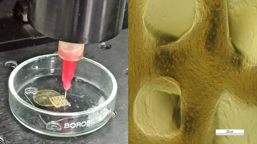
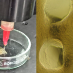
The researchers developed a 3D-printable silk fibroin-gelatin scaffold linked to LDN-193189, a small molecule that blocks bone morphogenetic protein (BMP) signalling. Earlier studies have shown that excessive BMP signalling drives cartilage degeneration and promotes osteoarthritis.
“Our earlier studies showed that excessive BMP signalling pushes cartilage cells towards bone formation and worsens osteoarthritis. We also found that blocking this pathway helps regenerate healthy cartilage while reducing inflammation,” said Prof. Bandyopadhyay.
The implant combines two biological strategies. The silk scaffold encourages cartilage formation through Wnt/β-catenin signalling, while the attached drug (LDN-193189) blocks BMP signalling, which is linked to cartilage degeneration. “We chemically attached the drug to the scaffold instead of simply loading it into the material. This allowed the drug to remain active for longer in the damaged joint,” said Sayeda Fauzia Iqbal, lead author of the study.
To test the implant, the team first induced osteoarthritis in rats by cutting the anterior cruciate ligament. A full-thickness cartilage defect was then created in the knee. This model closely resembles the diseased environment seen in human osteoarthritis.
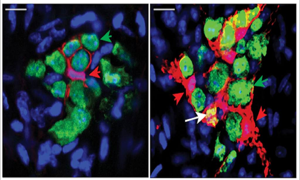
Four months after implantation, the treated animals showed extensive cartilage regeneration. Tissue analysis showed strong Safranin O staining, indicating restoration of cartilage proteoglycans. The regenerated tissue also expressed collagen type II, a marker of healthy hyaline cartilage. At the same time, collagen type I and collagen type X, markers of fibrocartilage and hypertrophic cartilage, were largely absent.
Besides promoting cartilage growth, the implant also reduced inflammation. The researchers found no detectable expression of NF-κB, a key regulator of inflammatory signalling in osteoarthritis, in the regenerated tissue after four months.
The study also produced an unexpected result. The researchers initially expected that scaffolds carrying living cells would perform better. Instead, the cell-free scaffold regenerated cartilage more effectively. The findings suggest that the scaffold recruited the body’s own cells to repair the tissue when given the right biological signals and mechanical support.
He said the main strength of the work was its dual approach. “The scaffold suppresses abnormal BMP signalling while naturally activating Wnt/β-catenin signalling. This is more sophisticated than simply delivering a growth factor. Covalently attaching LDN-193189 also solves its short half-life problem,” he said.
He cautioned that the work remains at an early stage. Rat joints do not experience the same mechanical loads as human joints. The technology must therefore be tested in larger animals before clinical trials.
The researchers plan to carry out safety studies and evaluate the scaffold in larger animals. If the approach succeeds in larger animal studies and human trials, it could eventually offer patients a way to repair damaged cartilage instead of relying solely on pain relief or joint replacement surgery.
Disclaimer:
SciSoup claims no competing interest. To ensure accuracy and scientific relevance, this science blog has been reviewed by the research team involved in the study.


“We wanted to develop a biomaterial that could promote cartilage healing while ensuring only hyaline cartilage formation in an osteoarthritic environment,” said Prof. Amitabha Bandyopadhyay, who led the study at IIT Kanpur.